Downloaded 183 times


![Feature: Spiculation
[Huo et al.]
• Extract the mass using a
region-growing technique
• The maximum gradient and
its angle relative to the
radial direction are
computed
• Calculate the full-width at
half-maximum (FWHM)
from the cumulative
gradient orientation
histogram
94](/p?url=https%3A%2F%2Fimage.slidesharecdn.com%2Fperjeta-140112203001-phpapp02%2F85%2FPerjeta-94-320.jpg&__src=https%3A%2F%2Fwww.slideshare.net%2Fslideshow%2Fperjeta%2F29942780&__type=image)
![Feature: Spiculation [Chan et al.]
• Determine the outline of
the segmented mass
• Obtain the rubber-bandstraightening-transformed
image
– The spicules become
approximately aligned in a
similar direction
• The rectangular region can
then be subjected to
texture analysis
95](/p?url=https%3A%2F%2Fimage.slidesharecdn.com%2Fperjeta-140112203001-phpapp02%2F85%2FPerjeta-95-320.jpg&__src=https%3A%2F%2Fwww.slideshare.net%2Fslideshow%2Fperjeta%2F29942780&__type=image)


![A Mammography CAD System
[Giger et al.]
Probability of
malignancy
Similar images of
known diagnosis
Indicates the unknown
lesion relative to all
lesions in the database
99](/p?url=https%3A%2F%2Fimage.slidesharecdn.com%2Fperjeta-140112203001-phpapp02%2F85%2FPerjeta-99-320.jpg&__src=https%3A%2F%2Fwww.slideshare.net%2Fslideshow%2Fperjeta%2F29942780&__type=image)

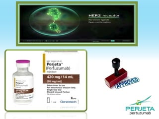
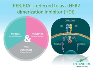
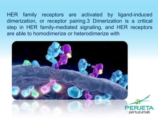
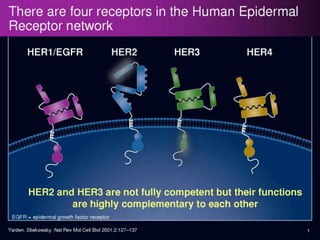
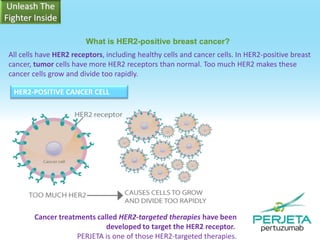
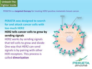
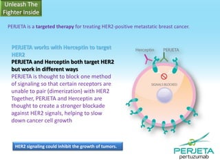
Perjeta® is an FDA-approved first-line treatment for HER2-positive metastatic breast cancer, used in combination with Herceptin (trastuzumab) and docetaxel (chemotherapy). This targeted therapy blocks HER2 signaling, helping to control cancer growth, and has been shown to significantly increase the time patients live without disease progression and improve tumor shrinkage rates. The combination of Perjeta, Herceptin, and docetaxel enhances the efficacy of treatment compared to Herceptin and docetaxel alone.


