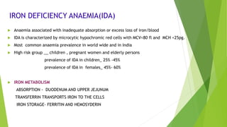
ANAEMIA-
IRON DEFICIENCY ANAEMIA
ASWATHY.A
ASSISTANT PROFESSOR
DEPARTMENT OF PATHOLOGY
DEFINITION
Anaemia refer to a decrese in the total number of circulatory red cells with decrease in HB,PCV
below the previously established normal values , health person of same age group ,gender race,and
similar environmental conditions.
Its clinical diagnosis is made from the history , physical examination ,sign and symptoms ,HB values and
other procedures and findings
IRON DEFICIENCY ANAEMIA(IDA)
 Anaemia associated with inadequate absorption or excess loss of iron/blood
 IDA is characterized by microcytic hypochromic red cells with MCV<80 fl and MCH <25pg.
 Most common anaemia prevalence in world wide and in India
 High risk group __ children , pregnant women and elderly persons
prevalence of IDA in children_ 25% -45%
prevalence of IDA in females_ 45%- 60%
 IRON METABOLISM
ABSORPTION - DUODENUM AND UPPER JEJUNUM
TRANSFERRIN TRANSPORTS IRON TO THE CELLS
IRON STORAGE- FERRITIN AND HEMOSYDERIN
DAILY REQUIREMENT OF IRON
Diet should contain 10-15 mg of elementary iron and with approximately 8-10% absorbed
Daily net requirement of males- 1 mg
Daily net requirement of females – 1.5 mg
FACTORS PROMOTING IRON ABSORPTION
Hcl of stomach
Ascorbic acid
FACTORS HAMPERING IRON ABSORPTION
Phytates of cereals
Tannate of tea
Phosphate of diet and drugs
Milk
Small loss of iron each day in urine, faeces,skin and nails and in menstruating females as blood (1-2 mg daily)
CAUSES
1. Decreased supply
2. Impared absorption
3. Increased demand
4. Loss
1) DECREASED SUPPLY
a) Nutritional deficiency
b) Malabsorption
2) IMPARED ABSORPTION
Total or partial gastrectomy impairs iron absorption by decreasing Hcl and transit time through the
duodenum
3)INCREASED DEMAND
increased utalisation ,
a) pregnancy and lactation
b) Growth- growing infants ,childrens and adolescents
4)LOSS
a) chronic blood loss - Gastro intestinal tract, urinary tract, genital tract and respiratory tract bleeding
b) Intravascular hemolysis – PNH, microangiopathic and hemolytic anaemia
CLINICAL FEATURES
 CRACKS IN THE SIDE OF THE MOUTH
 EXTREME FATIGUE (TIREDNESS)
 CHEST PAIN
 DIZZINESS / LIGHT HEADACHE
 GLOSSITIS
 Dysphagia( Plummer-Vinson syndrome)
 ATROPIC GASTRITIS
 PALE SKIN
 SPOON SHAPED NAILS, KOILONYCHIA,

 HAIR LOSS
 BLUE SCLERAE
 PICA (APETITE FOR NON FOOD SUBSTANCESSUCH AS AN ICE, CLAY)
 ANGULAR STOMATITIS
 IMMUNE FUNCTION
 BEETURIA
 SPLENOMEGALY (10%)
 CONGENITAL HEART FAILURE
LAB DIAGNOSIS OF IDA
1) Peripheral blood findings
2) Bone marrow examination
3) Iron state
PERIPHERAL BLOOD FINDINGS
a)Counting
HB-Decreased
PCV-Decreased
RBC- Normal
TLC-Normal
DC- Normal
Platelet- Increased
Red cell indices - MCV < 80fl
MCH< 25pg
MCHC< 27g/dl
RDW -Increased
Recticulocyte count-Normal or Increased
PERIPHERAL SMEAR EXAMINATION
RBC- Microcytic hypochromic anaemia with pencil/cigar shaped cells
Hypochromia- Central pallor being more than 1/3 rd
WBC- Normal size and shape
PLATELET- Increased in number and seen in groups
Severe anaemia – Central pallor 2/3 rd -3/4 th
BONE MARROW EXAMINATION
 Bone marrow is hypercellular
 Erythroid hyperplasia is present but is less as compared to the degree of anaemia .It varies from 2:1 to 1:2
 Miconormoblastic reactions
 Myelopoiesis and megakaryopoiesis is normal
 Depletion of bone marrow iron-Prussion blue stain
Iron granules present in perls prussion blue stain
ASSESSMENT OF IRON STATE
 S. Ferritin- <12 µg/L
 S. Iron –reduced 10-15µg/dl
 TIBC- reduced (350-450µg/dl)
 Transferrin saturation- <16%
 STRA- Increased
 Red cell protoporphyrin- increased ( >200 g/dl)
 Recticulocyte hemoglobin content – reduced
 Erythrocyte zinc protoporphyrin – increased
DIFFERENTIAL DIAGNOSIS
 Thalassemia major and minor
 Anemia of chronic disorders
 Sideroblastic anemia
 Hb E thalassemia
 Lead poisoning
TREATMENT
Before using iron medications check if you are allergic to any drugs or food dyes or if you have,
 Iron overload syndrome
 Hemolytic anemia (a lack of red blood cells)
 Porphyria (a genetic enzyme disorder that causes symptoms affecting the skin or nervous system thalassemia
(a genetic disorder of red blood cells)
 Liver or kidney disease
 If you are an alcoholic; or if you receive regular blood transfusion
1) Oral iron therapy
2) Parenternal iron therapy
3)Non pharmacological treatment
ORAL IRON THERAPY
Oral iron treatment may require 3-6 months to replenish body iron stores.
 Ferrous sulfate is the DOC for iron deficiency anemia.
 Dosage: 325 mg, which provides 180 mg of iron daily of which 10mg is
usually absorbed.
 Patients who cannot tolerate iron on an empty
stomach should take it with food.
COMMON ADVERSE EFFECTS OF ORAL IRON THERAPY
• Nausea
• Epigastric discomfort
• Abdominal cramps
• Constipation and diarrhea.
• Black stool
• These effects are usually dose-related
PARENTERNAL IRON THERAPY
Indicates in,
 Late stage of pregnancy
 Post operative patients
 Patients who are unable to take oral preparation
 Iron Sorbitol is given as a single dose/weekly/daily
 Iron Dextran -Is a stable complex of ferric hydroxide and low-molecular weight dextran containing 50mg
of elemental iron per milliliter of solution . It can be given deep IM injection or IV infusion’
Adverse effect:
 light-headedness, fever, arthralgias, back
 pain, urticaria, bronchospasm and
 hypersensitivity reaction
NON PHARMACOLOGICAL TREATMENT
 Iron-rich diet
 Good sources of iron includes:
 Meats - beef, pork, lamb, liver, and other organ meats
 Poultry - chicken, duck, turkey, liver (especially dark meat)
 Fish - shellfish, including clams, mussels, and oysters, sardines, anchovies
 Leafy greens of the cabbage family, such as broccoli , kale, turnip greens, and collards
 Legumes, such as lima beans and green peas; dry beans and peas, such as pinto beans, black-eyed peas,
and canned baked beans
Iron Deficiency Anaemia